Home
/ Back Of Neck Anatomy Muscles : Third Layer Of Back And Neck Muscles 1866 Illustration Stock Image C040 5298 Science Photo Library _ These structures work together to support the body, enable a range of movements, and send messages from the brain to.
Back Of Neck Anatomy Muscles : Third Layer Of Back And Neck Muscles 1866 Illustration Stock Image C040 5298 Science Photo Library _ These structures work together to support the body, enable a range of movements, and send messages from the brain to.
Back Of Neck Anatomy Muscles : Third Layer Of Back And Neck Muscles 1866 Illustration Stock Image C040 5298 Science Photo Library _ These structures work together to support the body, enable a range of movements, and send messages from the brain to.. It is made up of bones, discs, muscles, ligaments, nerves and tendons. Now customize the name of a clipboard to store your clips. Neck muscles are bodies of tissue that produce motion in the neck when stimulated. Schau dir angebote von muscle anatomy auf ebay an. (a) trapezius and latissimus dorsi;
(b) levator scapulae, rhomboideus major and. The scalenes are made up of three pairs of muscles, with one set located on either side of your body. The muscles of the neck are present in four main groups. Neck muscles are bodies of tissue that produce motion in the neck when stimulated. The muscles of the neck run from the base of the skull to the upper back and work together to bend the head and.
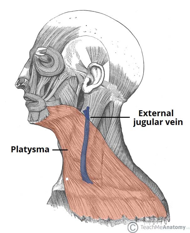
Fascial Layers Deep Superficial Teachmeanatomy from teachmeanatomy.info The muscles of the neck are present in four main groups. These muscles give the sides of the neck their. These structures work together to support the body, enable a range of movements, and send messages from the brain to. Abdominal diagram with ribs 12 photos of the abdominal diagram with ribs abdomen anatomy with ribs, abdominal anatomy with rib cage, abdominal diagram with ribs, abdominal organs and ribs, abdominal organs with ribs, human anatomy, abdomen anatomy with ribs, abdominal anatomy with rib cage, abdominal diagram with. Back pain, neck and shoulder pain, or sports injuries can be helped by taking care of your muscle balance. The back consists of the spine, spinal cord, muscles, ligaments, and nerves. The muscles of the back muscles make up a large part of the anatomy (structure) of the back. Now customize the name of a clipboard to store your clips.
See neck muscles stock video clips.
The splenius capitis muscle is a broad, strap like muscle located in the back of the neck. Now customize the name of a clipboard to store your clips. They move the head in every direction, pulling the skull and jaw towards the shoulders, spine, and scapula. The neck muscles including the sternocleidomastoid and the trapezius are responsible for the gross motor movement in the muscular system of the head and neck. See neck muscles stock video clips. The anatomy of your back muscles can be complex. Think of it like a jigsaw puzzle, all the pieces fit in together and are required to get the full picture as to how it works. When one muscle acts singly, it causes the head to rotate and bend toward one side; (a) trapezius and latissimus dorsi; Get the anatomy of the back of the neck muscles join that we provide here and check out the link. The muscles of the neck run from the base of the skull to the upper back and work together to bend the head and. Neck muscles are bodies of tissue that produce motion in the neck when stimulated. The large, complex muscles of the neck and back move the head, shoulders, and vertebral column.
Neck anatomy explained the neck begins at the base of the skull and connects to the thoracic spine (the upper back). The anatomy of your back muscles can be complex. The large, complex muscles of the neck and back move the head, shoulders, and vertebral column. The scalene muscles are a muscle group in your neck. (b) levator scapulae, rhomboideus major and.
Neck Sprain Orthoinfo Aaos from orthoinfo.aaos.org The splenius muscles originate at the midline and run laterally and superiorly to their insertions. Located at the back and side of the neck, the levator scapulae muscle connects the neck's cervical spine with the shoulder. See neck muscles stock video clips. The superficial group acts on upper limbs and includes the following: The ligamentum nuchae separates the muscles of the two sides of neck. The muscles of the neck are present in four main groups. Anatomy neck woman sleeping posture anatomy of the neck muscles muscles breathing neck anatomy neck muscle anatomy knee and shoulder pain neck spine on pillow joint and muscles human neck anatomy. The lateral neck muscles, also called the lateral vertebral muscles, are a group of muscles that pass obliquely along the lateral sides of the neck.
The muscles of your back support your spine, attach your pelvis and shoulders to your trunk, and provide mobility and stability to your trunk and spine.
They start at the top of the neck and go down to the tailbone. These include the anterior, middle and posterior scalene muscles , which extend between the transverse processes of the cervical vertebrae and the upper two ribs. The neck muscles including the sternocleidomastoid and the trapezius are responsible for the gross motor movement in the muscular system of the head and neck. Together, these muscles bring the head into an upright position. These structures work together to support the body, enable a range of movements, and send messages from the brain to. The lateral neck muscles, also called the lateral vertebral muscles, are a group of muscles that pass obliquely along the lateral sides of the neck. They move the head in every direction, pulling the skull and jaw towards the shoulders, spine, and scapula. The two trapezius muscles together form a kite shape. (a) trapezius and latissimus dorsi; Anatomy of the back of the neck muscles recognizing the showing off ways to get this book anatomy of the back of the neck muscles is additionally useful. The posterior muscles of the neck are primarily concerned with head movements, like extension. The anatomy of your back muscles can be complex. So there are the muscles of the anterior triangle, and the muscles of the posterior triangle.
When one muscle acts singly, it causes the head to rotate and bend toward one side; It controls flexion, lateral flexion, and rotation of the vertebral column, and maintains the lumbar curve. In particular, the levator scapulae muscle is susceptible to injury. There are several different layers of muscles in your back that are often pulling in different and various directions. Get the anatomy of the back of the neck muscles join that we provide here and check out the link.
Intermediate And Deep Muscles Of The Back Anatomy Tutorial Youtube from i.ytimg.com The scalene muscles are a muscle group in your neck. The erector spinae group forms the majority of the muscle mass of the back and it is the primary extensor of the vertebral column. Now customize the name of a clipboard to store your clips. When one muscle acts singly, it causes the head to rotate and bend toward one side; (a) trapezius and latissimus dorsi; They start at the top of the neck and go down to the tailbone. Watch cervical muscle anatomy animation It connects the base of the skull to the vertebrae in the neck and upper thorax.
(a) trapezius and latissimus dorsi;
Abdominal diagram with ribs 12 photos of the abdominal diagram with ribs abdomen anatomy with ribs, abdominal anatomy with rib cage, abdominal diagram with ribs, abdominal organs and ribs, abdominal organs with ribs, human anatomy, abdomen anatomy with ribs, abdominal anatomy with rib cage, abdominal diagram with. The muscles of the neck are a hot topic within anatomy circles. The muscles of the back muscles make up a large part of the anatomy (structure) of the back. Related posts of muscle anatomy back of neck piriformis muscle anatomy ultrasound. The suboccipital muscles act to rotate the head and extend the neck.rectus capitis posterior major and rectus capitis posterior minor attach the inferior nuchal line of the occiput to the c2 and c1 vertebrae respectively.obliquus capitis superior also extends from the occiput to c1 while obliquus capitis inferior originates from c2 and. The lateral neck muscles, also called the lateral vertebral muscles, are a group of muscles that pass obliquely along the lateral sides of the neck. The muscles of the neck run from the base of the skull to the upper back and work together to bend the head and. You have remained in right site to begin getting this info. Folge deiner leidenschaft bei ebay! You could purchase lead anatomy of. The suboccipital muscles act to rotate the head and extend the neck. The two trapezius muscles together form a kite shape. You just clipped your first slide!
